
X-rays show what remains hidden to the naked eye during an examination: decay between the teeth, inflammation at the root tip, the condition of the jawbone or the position of wisdom teeth. At our practice in Küsnacht, we take digital X-rays in 2D and, when needed, in 3D. We only take images that are necessary for your diagnosis or treatment, and we choose the method with the lowest radiation exposure that can answer the question.
For digital impressions and documentation, we also use an intraoral scanner, which works entirely without X-rays. We explain the difference further down and in detail in our article 3D scanning in dentistry.
Types of X-ray
Single and bitewing X-rays
For these small images, the image receptor is placed inside your mouth. A single X-ray shows one or a few teeth, including the root and surrounding bone, for example if inflammation at the root tip is suspected or during root canal treatment. A bitewing X-ray shows the back teeth of the upper and lower jaw at the same time. It mainly helps to detect decay between the teeth, which is often not visible from the outside.
For these images, we use the VistaScan digital phosphor plate system from Dürr Dental. The thin, flexible plate is scanned after the exposure, and shortly afterwards we can look at the image on screen together with you.
Panoramic X-ray (OPG)
The panoramic X-ray, also called an orthopantomogram or OPG, shows all the teeth, both jaws, the jaw joints and parts of the maxillary sinuses in a single image. It provides an overview, for example during a comprehensive initial examination, before wisdom teeth are removed or in orthodontics. For fine details of individual teeth, the small images are more suitable.
Cephalometric X-ray
The cephalometric X-ray is a side view of the skull. It shows how the upper and lower jaws are positioned relative to each other and how the front teeth sit in the bone. In orthodontics, angles and distances are measured on this image. This helps with planning and, in children and adolescents, with assessing jaw growth.
3D X-ray: cone beam computed tomography (CBCT)
CBCT (cone beam CT), known in German as DVT, produces a three-dimensional image of a section of the jaw. Bone, tooth roots, the nerve canal and the maxillary sinus can be viewed from all directions without overlapping structures. Because the radiation dose is higher than with 2D images, we use it selectively.
Our unit: Orthophos SL 3D with cephalometry
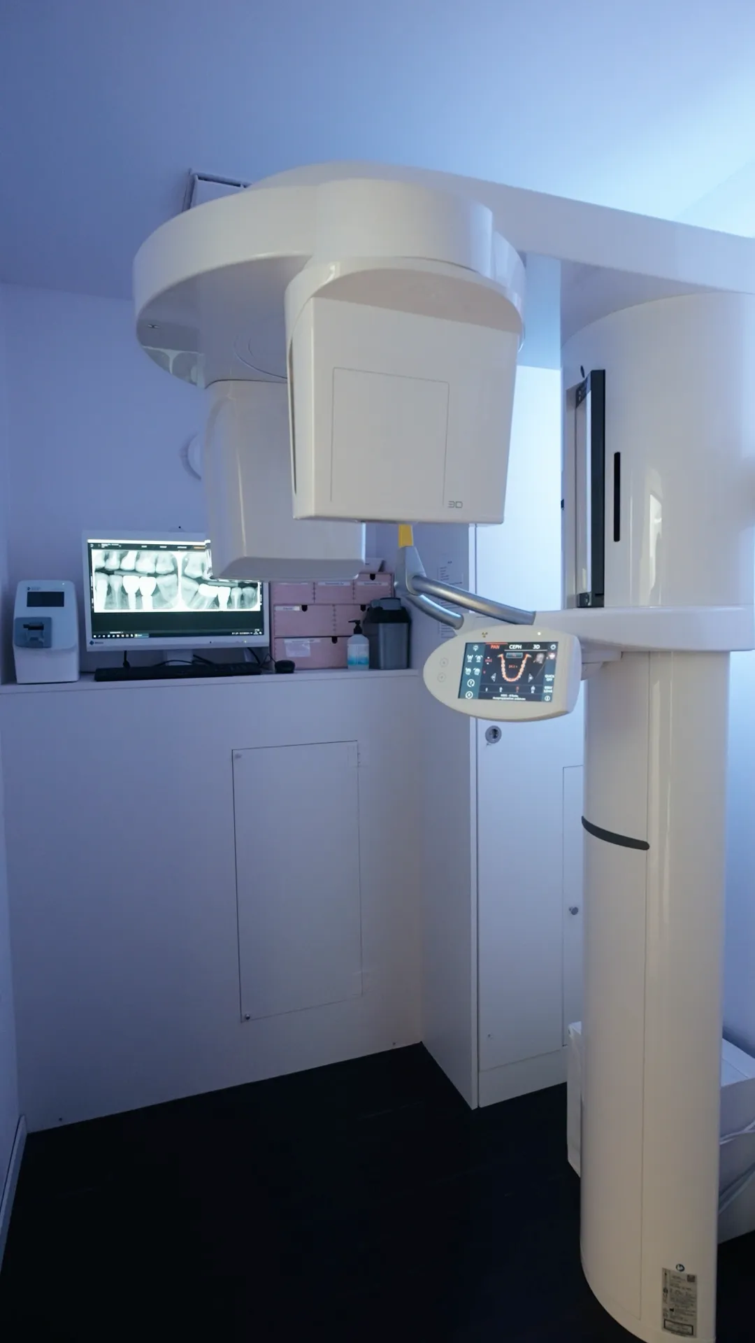
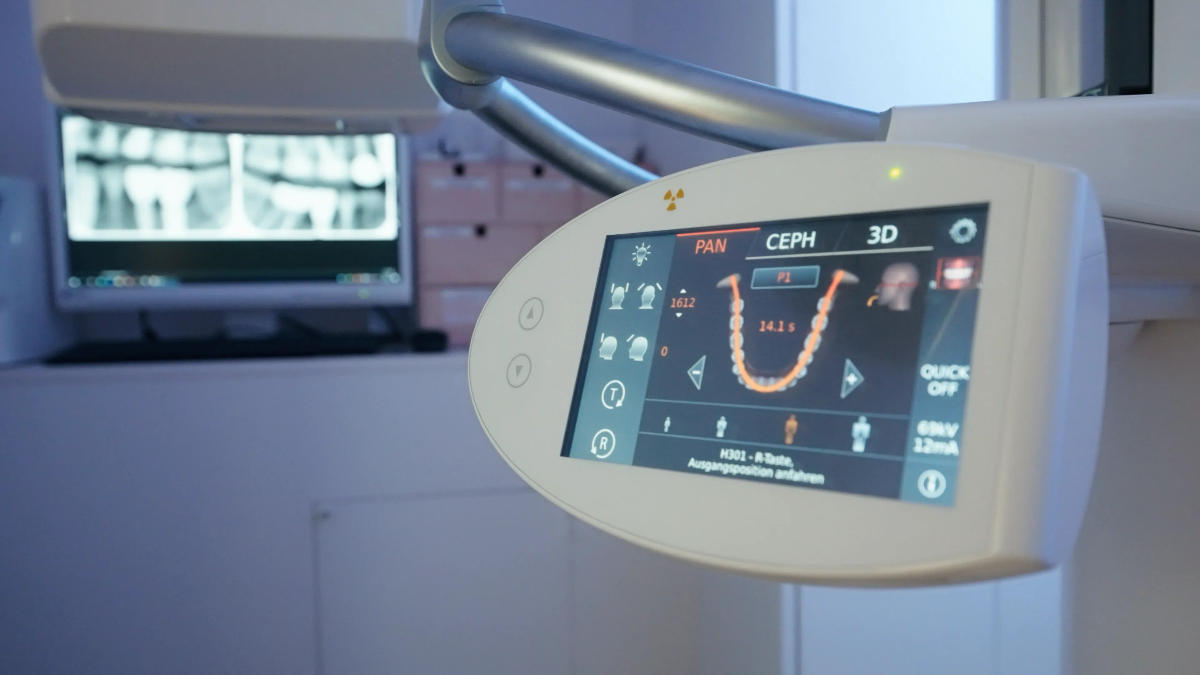
For the larger images, we use the Orthophos SL 3D from Dentsply Sirona. The unit combines three methods: panoramic X-rays (2D), three-dimensional images (CBCT) and, thanks to the additional cephalometric arm, cephalometric X-rays.
With 3D images, the size of the imaged area can be selected. This is important for radiation protection: according to the Federal Office of Public Health (FOPH), the simplest way to lower the dose of a CBCT scan is to reduce the volume imaged. We therefore only capture the area needed to answer the clinical question.
During the scan, you bite on a small bite block while side supports keep your head still; the exposure itself takes only seconds.
When a 3D scan (CBCT) makes sense
A CBCT scan is not a routine examination. It is indicated when an examination and 2D images are not enough to plan treatment safely. Typical situations include:
- Implant planning: The 3D image shows the height and width of the bone and the distance to the nerve and maxillary sinus. For dental implants, we combine it with the intraoral scan and use both to plan a surgical guide for guided implant placement.
- Wisdom teeth: If the panoramic X-ray suggests that a lower wisdom tooth lies very close to the mandibular nerve, a CBCT scan clarifies its exact position before wisdom tooth removal. Without such signs, a 2D image is usually sufficient.
- Complex root canal treatments: for example with unclear symptoms, canals that are hard to locate or severely curved, or a suspected root fracture, when 2D images do not provide an answer.
- Impacted teeth: for example a canine that does not erupt.
- Bone loss around a tooth: in individual cases during the treatment of gum and bone disease, to show bone pockets in more detail.
For children and adolescents, the indication is particularly strict; CBCT is not recommended for routine orthodontic diagnosis.
Radiation protection and safety
In Switzerland, the operation of X-ray equipment is governed by the Radiological Protection Ordinance and the X-ray Ordinance, with requirements set by the Federal Office of Public Health (FOPH). Three principles apply:
- Justification: An image is only taken if its benefit for your diagnosis or treatment outweighs the radiation risk.
- Optimisation: Any unnecessary dose is avoided, in line with the internationally recognised ALARA principle (‘as low as reasonably achievable’).
- Training: Dentists need training in radiation protection and must complete further training at least every five years. The FOPH requires additional training for CBCT.
For context: the FOPH puts the effective dose of a small dental X-ray at around 5 microsieverts and that of a CBCT scan at around 100 microsieverts, roughly twenty times as much. Small dental X-rays are among the medical X-ray examinations with the lowest dose. Even so, the rule is: as much as necessary, as little as possible.
Regular maintenance and inspection
Our X-ray units are serviced and inspected every year in accordance with FOPH requirements. The FOPH guideline for dental X-ray systems stipulates that a specialist company tests the system after servicing (status test). In between, test phantoms are used regularly to check that image quality and dose remain unchanged (constancy test). All results are recorded.
Lead apron and thyroid shield
For adults, a lead apron is no longer standard for dental X-rays. According to the joint 2023 recommendation of the Swiss Society of Dentomaxillofacial Radiology (SGDMFR) and the SSO, it offers no significant additional benefit at today’s low doses. For children and adolescents until they have finished growing, the recommendation still provides for a thyroid shield: for small X-rays, for cephalometric X-rays and, depending on the area imaged, for CBCT.
X-ray and 3D scan: the difference
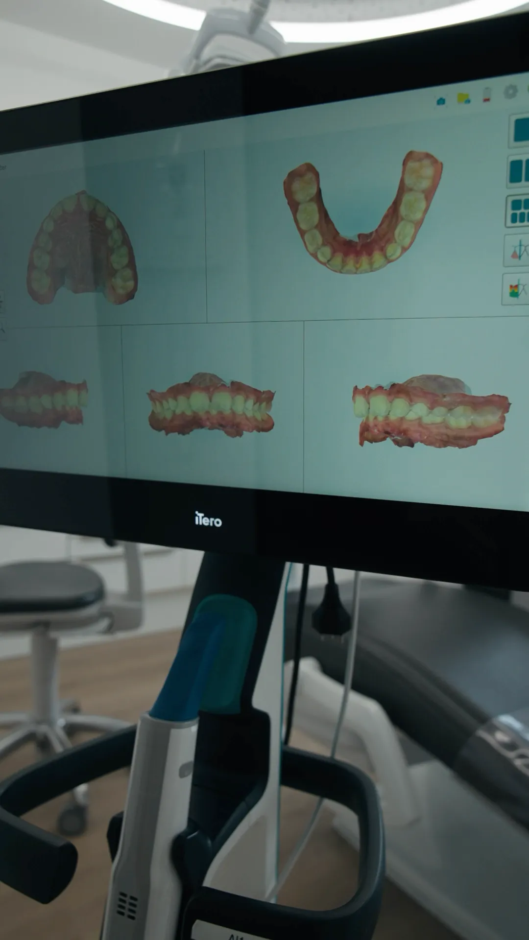
Our iTero intraoral scanner works with light, not X-rays. It captures the surface of the teeth and gums and uses this to create a digital 3D model that replaces the traditional impression. We use it for planning, for example for Invisalign, and routinely for documentation.
The scan does not show what lies beneath the surface: we can only see decay inside the tooth, roots, inflammation in the bone or the course of nerves on an X-ray. The two methods complement each other, for example in implant planning.
Frequently asked questions
Can I have an X-ray during pregnancy?
Please tell us before any X-ray if you are or might be pregnant. With dental X-rays, the abdomen is not in the radiation field, and the dose to the unborn child is very low. Even so, we weigh things up particularly carefully during pregnancy: where possible, we postpone X-rays that are not urgent until after the birth. If you have acute symptoms, a targeted X-ray may be advisable.
How often are X-rays taken?
There is no fixed schedule. Whether and when a new X-ray is needed depends on your situation, for example on your risk of decay, previous findings or planned treatment. You are welcome to bring recent X-rays from another practice; this often avoids duplicate images.
Do you also X-ray children?
Yes, if it is necessary for the diagnosis, for example if decay between the milk teeth is suspected, after a dental injury or before orthodontic treatment. For children, the indication is assessed particularly strictly, and depending on the type of image, the recommendations provide for a thyroid shield. You can read more about treating our youngest patients under Children and family.
We are happy to explain why we recommend an X-ray and what it shows. Book your appointment online here or call us on +41 44 301 26 02.
Appointments & advice
Questions, or ready to book an appointment?
Write to us and we will get back to you as soon as possible. Prefer to book online or give us a call? That works too.
Karamousli Dental Practice · Seestrasse 137 · 8700 Küsnacht